Oral Cancer Screenings in Pineville, Charlotte: Why Early Evaluation Matters
Short answer: A persistent mouth sore, lump, or unexplained patch should be evaluated. An examination may identify concerns, but further testing is needed to diagnose cancer. National Institute of Dental and Craniofacial Research.
An oral cancer examination can help identify suspicious changes in the mouth and throat. Screening does not diagnose cancer; an abnormal finding may require further evaluation and a tissue biopsy. A sore, lump, or red or white patch lasting more than two weeks deserves prompt assessment.
Tobacco and heavy alcohol use increase oral cancer risk. Tell your dental team about tobacco use and any persistent changes in your mouth. A suspicious-looking patch is not itself a cancer diagnosis.
Quick Insights: Oral Cancer Screenings
- Oral cancer can develop without pain, making routine dental screenings important for detecting suspicious changes early.
- Know the warning signs: mouth sores lasting more than two weeks, persistent white or red patches, lumps, unexplained bleeding, numbness, difficulty swallowing, or ongoing hoarseness.
- Know your risk: tobacco, heavy alcohol use, HPV, prolonged sun exposure, age, and a previous oral cancer diagnosis can increase risk.
- Screening is quick and systematic, typically involving a visual and physical examination of the mouth, tongue, throat, jaw, and neck. Advanced tools may help evaluate suspicious areas.
- Suspicious findings may require further testing, such as a brush biopsy, surgical biopsy, or imaging, to determine whether abnormal tissue is cancerous.
- Regular dental visits provide an opportunity for early detection, while high-risk patients or those with persistent lesions may need more frequent monitoring.
- Prevention matters: avoiding tobacco, limiting alcohol, protecting your lips from UV exposure, and getting recommended HPV vaccination can help reduce risk.
Understanding Oral Cancer Risk
Tobacco, heavy alcohol use, age, and some HPV infections are relevant risk factors. Risk differs by cancer site and individual history. Having a risk factor does not establish a diagnosis, and people without typical risk factors can still develop cancer.
Screening Process and Techniques
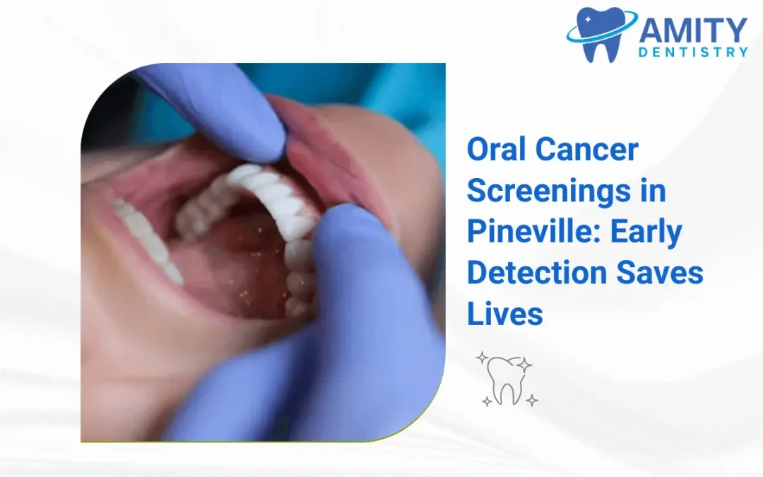
Visual examination forms the foundation of oral cancer screening. Dentists inspect all visible oral tissues under bright lighting. Systematic evaluation covers lips, gingiva, buccal mucosa, tongue surfaces, floor of mouth, hard and soft palates, and oropharynx.
Practitioners look for asymmetries, color variations, ulcerations, and unusual growths. Red patches called erythroplakia and white patches termed leukoplakia warrant close attention. Texture changes including roughness, firmness, or induration indicate potential problems.
Palpation follows visual inspection. Dentists use gloved hands to feel for lumps, masses, or abnormal tissue consistency. Bimanual examination of the floor of mouth and tongue base detects deeper abnormalities. Neck palpation identifies enlarged lymph nodes suggesting metastatic spread.
Adjunctive screening tools supplement standard examination. VELscope technology uses blue light fluorescence to highlight abnormal tissue. Suspicious areas appear darker than surrounding healthy tissue. Brush biopsy techniques collect cell samples from questionable lesions without surgical intervention.
Toluidine blue staining marks dysplastic and cancerous cells. Providers apply the dye to suspicious areas and rinse after one minute. Abnormal tissue retains the blue color while normal tissue clears. Positive staining requires biopsy confirmation.
When to Discuss Oral Cancer Screening
Routine dental visits provide opportunities to examine the mouth. The timing of further checks depends on symptoms, risk factors, and any prior diagnosis. People with previous cancer or precancerous findings should follow their treating team’s plan.
Do not wait for a routine appointment if a sore, lump, red or white patch, or other unexplained change persists for more than two weeks. Difficulty breathing or swallowing needs immediate medical care.
Warning Signs and Symptoms
- Early oral cancer often produces no pain. Visible changes precede discomfort in most cases. A sore or irritation lasting more than two weeks represents the most common initial sign. These lesions fail to heal despite good oral hygiene.
- White or red patches appear on gums, tongue, tonsils, or mouth lining. Leukoplakia presents as thick white patches that cannot be scraped off. Erythroplakia shows as red velvety patches with higher malignant transformation rates than white lesions.
- Lumps or thickening in cheek tissues or neck indicate potential problems. Patients may notice difficulty moving the tongue or jaw. Chewing, swallowing, or speaking become uncomfortable or impaired. Dentures may suddenly fit poorly due to tissue changes.
- Persistent sore throat or hoarseness lasting more than two weeks requires investigation. Voice changes without upper respiratory infection suggest laryngeal involvement. Chronic cough unrelated to smoking cessation or respiratory illness warrants evaluation.
- Numbness or pain in mouth or lips develops as lesions progress. Ear pain occurs with advanced oropharyngeal cancers through referred nerve pathways. Unexplained bleeding from mouth or throat tissues signals tissue breakdown.
- Weight loss accompanies advanced disease when eating becomes difficult. Swollen lymph nodes in the neck persist beyond typical infection resolution timeframes. These nodes feel firm and fixed rather than mobile and tender.
Oral Cancer Assessment at a Dental Visit
A dental examination can include a visual and physical check of the mouth and surrounding tissues. Ask the Amity Dentistry team what assessment is appropriate for your symptoms and history. Suspicious findings may require referral for further evaluation.
Special lights, saliva tests, and brush tests are not substitutes for a tissue biopsy when one is indicated. A clinician should explain what a test can and cannot establish.
Diagnostic Follow-Up Procedures
Suspicious screening findings trigger diagnostic protocols. Incisional biopsy removes a portion of abnormal tissue for pathological examination. Excisional biopsy removes entire small lesions. Surgeons perform these procedures under local anesthesia in outpatient settings.
Fine needle aspiration evaluates neck masses and enlarged lymph nodes. Cytologists examine collected cells for malignant characteristics. This technique helps stage disease and plan treatment approaches.
Imaging studies determine cancer extent. Panoramic radiographs show bone involvement. CT scans detail soft tissue invasion and lymph node status. MRI provides superior soft tissue contrast for tongue and floor of mouth lesions. PET scans identify distant metastases in advanced cases.
Endoscopic examination visualizes the larynx, pharynx, and esophagus. Otolaryngologists perform these procedures to assess tumor extent and identify synchronous primary cancers. Approximately 15% of oral cancer patients develop second primary tumors in the upper aerodigestive tract.
Pathology reports grade tumors and identify histologic type. Staging incorporates tumor size, lymph node involvement, and metastatic spread. The TNM classification system guides treatment planning and prognosis estimation. Molecular testing identifies HPV status and other biomarkers affecting treatment decisions.
Treatment Options After Detection
Treatment is planned by the cancer care team according to the cancer’s site, stage, and the person’s health. Options may include surgery, radiation, and medicines. This article cannot determine which treatment is appropriate.
Radiation therapy treats cancers unsuitable for surgery or serves as adjuvant therapy after surgical resection. External beam radiation delivers targeted doses to tumor sites. Brachytherapy places radioactive sources directly into tumor tissue for concentrated treatment.
Advanced disease requires multimodal approaches. Surgery combined with radiation and chemotherapy improves outcomes for large tumors or cases with lymph node involvement. Chemotherapy uses drugs like cisplatin and 5-fluorouracil to kill cancer cells systemically.
Targeted therapy and immunotherapy represent newer treatment options. Cetuximab targets epidermal growth factor receptors on cancer cells. Pembrolizumab and nivolumab activate immune systems to fight cancer. These agents work alone or combined with traditional treatments.
Reconstructive surgery restores function and appearance after tumor removal. Tissue flaps from other body sites rebuild removed structures. Dental rehabilitation including implants and prosthetics restores chewing ability and speech clarity.
Why Prompt Evaluation Matters
Prompt evaluation of a persistent change can help identify its cause and avoid unnecessary delay. Screening cannot reliably predict an individual outcome. Prognosis depends on the diagnosis, stage, overall health, and response to treatment.
Reducing Risk
Avoid tobacco, discuss alcohol use with your health professional, and ask about HPV vaccination recommendations that apply to your age and history. Protect your lips from excessive sun exposure. These steps can reduce risk but cannot eliminate it.
Discuss a Concern with Amity Dentistry
Call (980) 423-1244 or request an appointment to discuss a persistent mouth change. The team will confirm availability and explain the next step. Specialist assessment may be needed.
Role of Dental Professionals
Dentists serve as frontline screeners given their regular patient contact. Biannual dental visits provide opportunities to detect cancer before symptoms develop. Professional training in oral pathology equips dentists to identify suspicious lesions.
Hygienists perform preliminary screenings during cleaning appointments. They document findings and alert dentists to abnormalities requiring further evaluation. This team approach maximizes detection sensitivity.
Patient education represents a critical professional responsibility. Dentists discuss risk factors and encourage behavior modification. Written materials and visual aids reinforce verbal counseling. Documentation of counseling satisfies medicolegal requirements and quality metrics.
Referral coordination ensures patients receive appropriate specialty care. Dentists maintain relationships with oral surgeons, otolaryngologists, and oncologists. Clear communication of clinical findings and preliminary impressions facilitates efficient diagnostic workup.
Survivorship care brings patients back to dental practices. Dentists manage oral complications of cancer treatment including xerostomia, mucositis, and radiation-related caries. Dental rehabilitation restores function after reconstructive surgery.
Related reading from Amity Dentistry
- The Importance of Regular Dental Checkups for Oral Health
- Signs You Need Dental Surgery: Key Symptoms to Watch
- Preventive Dentistry in Charlotte: How Regular Care Prevents Costly Dental Problems
Clinical content reviewed by Dr. Arati Shrestha, DMD on .
Sources
FAQs
How often should I get screened for oral cancer?
Discuss examination timing with your dentist. A persistent mouth change should be assessed without waiting for a routine visit. After a cancer diagnosis, follow the monitoring plan from your treating team.
Does oral cancer screening hurt?
A visual and gentle physical examination is usually well tolerated. Tell the clinician about soreness. If further testing is needed, ask what it involves and how discomfort is managed.
What happens if my dentist finds something suspicious?
Your dentist may recommend referral or further testing. A tissue biopsy may be needed to establish a diagnosis; a screening result alone does not confirm cancer.
Can oral cancer be cured if caught early?
Earlier diagnosis can provide more treatment options, but a cure cannot be promised. The cancer care team can explain prognosis for a specific diagnosis and stage.
Are oral cancer screenings covered by insurance?
Coverage depends on the exact plan and test. Ask the dental team and your insurer about benefits, authorization requirements, and any out-of-pocket cost.